Functional Magnetic Resonance Imaging (fMRI)
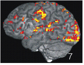
The following image shows brain activity measured during language processing. It was recorded with special fMRI techniques developed by Prof. Roland Beisteiner at the Medical University of Vienna.

Functional magnetic resonance imaging (fMRI) is a potent methodology to localize brain areas involved in or essential for specific brain functions. Recent studies related to the theoretical concept described above showed that the left pars opercularis increased its activation as a function of structural complexity of language (Makuuchi et al. 2009).
Menke et al. (2009) demonstrated that increased activity in the bilateral hippocampal formation, the bilateral fusiform gyri, the right precuneus and the right cingulate gyrus was associated with the degree of short-term treatment success with a certain form of aphasia therapy. In contrast, long-term training success was related to increased activity in the right-hemispheric Wernicke homologue.
A study investigating whistling activity in healthy subjects and oromandibular dystonia patients (Dresel et al. 2006) found differing brain activation changes in the primary motor cortex plus ventral premotor cortex (deficient activation) and the bilateral somatosensory areas plus supplementary motor area (overactivation) with dystonia patients.

University of Vienna
Althanstrasse 14
1090 Vienna


